Astaxanthin for eyes and skin

Astaxanthin for eyes and skin

March 24, 2025

Astaxanthin    Do you want to have beautiful, radiant skin and good health from the inside out? "Astaxanthin" is known as the "Queen of Antioxidants" with numerous benefits. What is Astaxanthin?   Astaxanthin is a nutrient in the carotenoid group, a red-orange substance that is extremely effective as an antioxidant. This has made Astaxanthin a very popular dietary supplement that helps nourish the skin, brighten the complexion, reduce wrinkles, nourish the eyes, and slow down cell degeneration from various pollutants. In addition to Astaxanthin having a powerful and excellent antioxidant role, it has a stronger antioxidant capacity than:6,000 times more than regular Vitamin C 800 times more than Co Q10 550 times more than Vitamin E 550 times more than Green tea catechins 75 times more than Alpha lipoic acid (ALA) 40 times more than Beta-carotene 17 times more than Grape seed extract  Where does astaxanthin come from?The human body cannot produce astaxanthin on its own; it must be obtained from food. It is most commonly found in most marine animals, such as salmon, trout, krill, shrimp, crayfish, and crabs. However, the most abundant natural source is the small red microalgae (Haematococcus pluvialis).     Who is Astaxanthin for?The body should receive 4–12 mg of Astaxanthin per day (pobpad.com).Astaxanthin has a unique structure that is both water-soluble and fat-soluble. The fat-soluble form is better absorbed by the body and is often found in softgel capsules.Normally, a person should take at least 4 mg per day, and a higher dosage is needed with age. However, if you want to focus on a specific benefit:For skin care: 4–18 mg/day. A higher dose is needed as you get older and skin deterioration increases to ensure it is effective.For eye care: 4–9 mg/day. It can be taken along with Lutein and Zeaxanthin.For high pollution exposure, such as from PM 2.5: 8–12 mg/day. This applies to people who are exposed to sunlight, pollution, or who play sports or work outdoors, such as motorcycle riders. Benefits of AstaxanthinThis antioxidant has many important benefits, including:Reducing inflammation in the body and strengthening the immune system.Slowing down the degeneration of the body in various systems, including blood, joints, tendons, muscles, the retina, the brain, and the nervous system.Helping to prevent dry and tired eyes, reducing eye pain, reducing free radicals in the eyes, increasing blood flow to the eyes, and preventing and restoring macular degeneration, which is common in the elderly and diabetic patients. It also helps protect the eyes from ultraviolet radiation.Protecting skin structure from damage by sunlight and ultraviolet radiation, helping to tighten pores, reduce wrinkles, and restore moisture and youthfulness.Enhancing the function of neurotransmitter cells in the brain, improving memory and reducing forgetfulness.Lowering blood pressure, increasing blood flow in capillaries, and helping to maintain normal blood lipid levels.Helping to increase muscle strength and helping the body recover faster.Preventing heart disease by protecting against inflammation in the blood vessels, which medical professionals point to as a major cause of heart disease today.Helping to strengthen the immune system, especially for patients with autoimmune diseases, low immunity, and chronic viral infections.Helping to prevent kidney and blood vessel degeneration in diabetic patients. ReferencesAstaxanthin, the most powerful antioxidant found in nature จากบล็อก https://www.yamamotonutrition.com/int/blog/post/astaxanthin-the-most-powerful-antioxidant-found-in-nature-a1754 Haematococcus pluvialis - Wikipedia จาก https://en.wikipedia.org/wiki/Haematococcus_pluvialis แอสต้าแซนธิน จาก https://www.thpherbal.com/product/22814-25764/แอสต้าแซนธิน Astaxanthin (แอสตาแซนทิน) - พบแพทย์ (pobpad.com) จาก https://www.pobpad.com/astaxanthin

EVIDENCE-BASED HEALTH BENEFITS OF COCONUT OIL

EVIDENCE-BASED HEALTH BENEFITS OF COCONUT OIL

March 23, 2025

EVIDENCE-BASED HEALTH BENEFITS OF COCONUT OILCoconut is one of the world's most significant tree crops, providing food and shelter to millions of people in the tropical region. Virgin coconut oil was introduced to the world at the end of twentieth century. Because of its wide range of applications in medication, food, cosmetics, and hair care products, virgin coconut oil is gaining worldwide popularity. Various methods have been employed to extract the coconut oil, but whichever method have been used it is still the best to avoid oil that has been refined, bleached or deodourized. This is because the health benefits, flavor, and scent of coconut oil are lost when it goes through these processes [1].Cold extraction and hot extraction are the two most used conventional methods of oil extraction. Low yield of oil is a challenge for these two procedures, and the heating process in the hot extraction approach reduces the antioxidant properties of the oil. The cold extraction method separates coconut oil from coconut milk without using any heat to break the emulsion. This technique has several advantages, including lower production costs because it does not require solvents or refinement operations like deodorizing and bleaching. As a result, the required amount of energy is reduced, making this procedure more environmentally friendly. Furthermore, when the coconut oil is refined, phytonutrients and polyphenols within the oil will be lost [1].Coconut oil comprises 90-95% of saturated fatty acids. Unlike long-chain fatty acids found in plant-based oil, medium-chain fatty acids are smaller in size, allowing higher cell permeability for immediate energy conversion instead of being stored as fat. Medium-chain fatty acids can also be digested more easily than long-chain fatty acids present in plant-based oil [1].At temperatures of 30°C and above, coconut oil is a colorless liquid. At a temperature of 25°C, it will solidify. Coconut oil contains of mainly saturated triglycerides, with medium chain acid which is lauric and myristic predominating. The main fatty acids in triglycerides oils are caprylic (C8) and capric (C10), which are both classed as medium-chain fatty acids, although lauric acid (C12), the main fatty acid in coconut oil, can be classified as either medium-chain fatty acids or long-chain fatty acids. Because the majority (70–75%) of lauric acid is absorbed with chylomicrons, it behaves more like a long-chain fatty acid in terms of digestion, whereas 95% of medium-chain fatty acids are absorbed directly into the portal vein. Medium-chain fatty acids are weak electrolytes and are highly ionized at neutral pH which increases their solubility. This marks the difference in solubility that occurs at chain lengths of C:10 and less, which excludes lauric acid [1]. HEALTH BENEFIT OF COCONUT OILInsulin Resistance, Inflammation, and ObesityObesity is characterized by an accumulation of adipose tissue either subcutaneously (underneath the skin) or ectopically (in and around organs). Certain fatty acids are more likely to be stored in adipose tissue rather than burned for energy when consumed. This can be measured by the fatty acid oxidation rate after fatty acid ingestion. When compared to certain long-chain saturated fats, medium-chain saturated fatty acids (C6-C12), such as lauric acid found in coconut oil, have relatively high oxidation rates [2].The overall evidence in the literature suggests that medium-chain saturated fatty acids is less likely to promote insulin resistance, inflammation, and fat storage compared to long-chain saturated fatty acids (such as stearic acid found in large quantities in butter, but particularly palmitic acid found in palm oil). While it is well known that fatty acids enter muscle tissue in order to be oxidized for ATP production used for muscle contraction, it is less commonly known that medium-chain saturated fatty acids may be directed more towards oxidation than long-chain saturated fatty acids. Thus, long-chain saturated fatty acids are more likely to be stored in muscle versus medium-chain saturated fatty acids, especially on top of a diet high in refined carbohydrates. This is why long-chain saturated fatty acids may cause greater muscle insulin resistance via increased intramyocellular lipid accumulation. Moreover, long-chain saturated fatty acids, as compared to unsaturated fats, are more readily incorporated into diacylglycerol (DAG) versus triacylglycerol (TAG), which may enhance inflammation and insulin resistance [2].In eight weeks of daily supplementation with coconut oil, Oliveira-de-Lira et al.  observed a higher adjuvant effect on weight loss and %body fat, with an emphasis on the reduction of anthropometric parameters associated with abdominal adiposity. Their findings are consistent with those reported by Assunção et al. and Cardoso et al., who reported a reduction in waist circumference that was associated with a significant increase in high-density lipoprotein cholesterol (HDL-C) after coconut oil supplementation as compared to placebo oil (a source of long-chain fatty acids) supplementation in obese women. Their findings regarding the reduction in waist circumference and conicity index reinforce the potential effects of coconut oil in causing a decrease in abdominal adiposity [3]. Coconut Oil on Blood Lipids Extra virgin coconut oil has recently been promoted as a healthy oil. Despite being high in saturated fat, the principal saturated fatty acid, lauric acid (C12), has been suggested to have different metabolic and hence health effects compared with other saturated fatty acids such as palmitic acid (C16), predominant in butter, palm oil and animal fat. In particular, it has been suggested that coconut oil does not raise total cholesterol (TC) or low density lipoprotein cholesterol (LDL-C) as much as butter [4].For a variety of reasons, the results were unexpected. Coconut oil is predominantly saturated fat which is generally held to have an adverse effect on blood lipids by increasing blood LDL-C concentrations. However, the saturated fatty acids profiles of various dietary fats differ significantly; coconut oil is predominantly (around 48%) lauric acid (C12) compared with butter (66% saturated fat) which is about 40% palmitic acid (C16) and stearic acid (C18), suggesting that coconut oil may not have the same health effects as other saturated fat-rich foods. Nevertheless, though reviews on coconut oil and cardiovascular disease risk factors have concluded that the evidence of an association between coconut oil consumption and blood lipids or cardiovascular risk was mostly poor quality, trials have generally reported that coconut oil consumption raises LDL-C in comparison to polyunsaturated oil such as safflower oil, though not as much in comparison to butter [4].Despite inconsistent findings in the literature, women who took coconut oil supplements had no negative alterations in their lipid profile. On the contrary, they experienced beneficial effects, such as an increased HDL-C levels and decreased triglycerides /HDL-C ratio as compared to the long-chain fatty acids group [3]. In addition, Nevin and Rajamohan  found that virgin coconut oil reduced total cholesterol, triglycerides, phospholoipids, low density lipoprotein (LDL), very-low-density lipoprotein (VLDL), and increased high density lipoprotein (HDL)-cholesterol levels. In vitro, LDL oxidation was reported to be prevented by the polyphenol component of virgin coconut oil [5].Other BenefitsVisna virus, Cytomegalovirus (CMV), Epstein-Barr virus, Influenza virus, Leukemia virus, Pneumonovirus, and Hepatitis C virus are all lipid-coated viruses that coconut oil is very effective against. Coconut oil's medium chain saturated fatty acids destroy these organisms by disrupting their membranes and interfering virus assembly and maturation. Monoglycerides are active against these viruses, but diglycerides and triglycerides are inactive. Lauric acid has more antiviral action than caprylic, capric, or myristic acids among the saturated fatty acids [5].Several authors have found that cold pressed oil has antimicrobial activity against Gram-positive and Gram-negative bacteria in vitro, in situ, and in vivo. Coconut oil exhibits antibacterial activity against Gram-positive and Gram-negative bacteria, according to Nevin and Rajamohan. Additionally, coconut oil has been used as a feed additive owing to its beneficial applications and health benefits such as antioxidant and anti-inflammatory activities [6].El-Abasy et al. found that coconut supplementation played a role in enhancing immune parameters and health. Furthermore, enhanced lysozyme activity with supplementation of coconut oil may indicate improved immunity in rabbits. More than 90% of the fatty acids in coconut oil are saturated and just less than 10% are unsaturated. Saturated fatty acids in coconut oil have been found to significantly enhance the innate immune responses [6].

THE ROLE OF CALCIUM AND VITAMIN D

THE ROLE OF CALCIUM AND VITAMIN D

March 22, 2025

THE ROLE OF CALCIUM AND VITAMIN DCalciumCalcium is the most abundant mineral in the body. Approximately 1.2 kg (equivalent to about 300 mmol) is contained within the human body, with 99% of this calcium being located within the bones and teeth. Calcium is also located in body fluids and soft tissues. It has two key roles: (1) supporting structural integrity; (2) regulating metabolic function. Cellular structure, intercellular and intracellular metabolism, signaling, heart muscle contractions, nerve function, enzyme activity, and normal blood clotting are all dependent on calcium. There is no functional marker of calcium status, since its role in normal blood clotting takes priority and hence plasma calcium is maintained within very narrow limits [1].The jejunum, ileum, and colon are the primary sites for calcium absorption. Uptake occurs by active transport and simple passive diffusion. Active transport is more prevalent when calcium intake is low, but when intake rises, more calcium is absorbed through non-specific routes. The metabolite of vitamin D (1,25-dihydroxycholecalciferol) stimulates calcium transport across the intestinal cells by inducing the production of a calcium-binding protein. This process occurs within the villus cells through the normal process of receptor binding, DNA interaction and messenger RNA production. Hence, vitamin D is critical for effective calcium absorption [1]. Vitamin D Vitamin D refers to vitamin D2 (ergocalciferol) or vitamin D3 (cholecalciferol). Ergocalciferol is produced from irradiated fungi or yeast. Cholecalciferol is produced in skin or found naturally in fatty fish such as salmon or mackerel. Although food can be fortified with both types of vitamin D, only cholecalciferol can be produced endogenously in skin. 7-dehydrocholesterol, a substance found in the skin, is converted into previtamin D3, which isomerizes to form vitamin D3, when exposed to ultraviolet B (UVB) radiation between the wavelengths of 290 and 315 nm. The amount of vitamin D3 made in the skin can be affected by an individual's skin color, age, and use of sunscreen products, as well as the time of day, season, and latitude [2]. Once vitamin D is made in the skin (D3) or obtained in the diet (D2 or D3), it enters the circulation bound to vitamin D–binding protein. The main form of vitamin D that circulates in the body is 25-hydroxyvitamin D (25(OH)D), which is created in the liver by the hydroxylation of vitamins D2 and D3 complex. The best marker for determining vitamin D level is 25(OH)D, which reflects both endogenous and exogenous sources. To generate the physiologically active form of vitamin D, 25(OH)D undergoes hydroxylation by the 1α-hydroxylase enzyme in the kidneys to produce 1,25-dihydroxyvitamin D (1,25(OH)2D; calcitriol). 1,25(OH)2D circulates bound to vitamin D–binding protein, enters the target cell, and binds to the vitamin D receptor (VDR) in the cytoplasm, which then enters the nucleus and heterodimerizes with the retinoic acid X receptor to increase transcription of vitamin D–dependent genes important for bone metabolism, calcium absorption, and other nonclassical functions (e.g., inhibition of genes important in cancer growth) [2, 3]. The Advantage of Calcium and Vitamin DOsteoporosisOsteoporosis is defined as a metabolic bone disease characterized by low bone mass and microarchitectural deterioration of bone tissue, leading to enhanced bone fragility and a consequent increase in fracture risk. According to estimates, three million people worldwide, roughly one in three women and one in twelve men aged > 50 years, will have osteoporosis at some point in their lives [1].Bone is a living tissue. Active bone formation (through the activity of osteocytes and osteoblasts) and bone resorption (involving osteoclasts) occur continuously. Osteoclasts are attracted to a quiescent bone surface and then excavate an erosion cavity. Mononuclear cells smooth off the erosion cavity, which is a subsequent site for the attraction of osteoblasts that synthesize an osteoid matrix. Continuous new bone matrix synthesis is followed by calcification of the newly-formed bone. When complete, lining cells once more overlie the trabecular surface [1]. It is well known that vitamin D promotes calcium absorption in the gut and kidney and helps to maintain adequate serum calcium concentrations to enable normal mineralization of the bone. Osteoblasts and osteoclasts need vitamin D to develop bones and repair them [4]. Bone matrix formation and bone maturation are stimulated by vitamin D. Additionally, it enhances osteoclastic activity and some evidence points to the possibility that it may influence differentiation of bone cell precursors [1]. Although vitamin D is utilized to enhance bone health, there is currently limited and conflicting evidence that vitamin D supplements alone have an impact on fracture outcomes. Thus, calcium and vitamin D work together synergistically on the bone [4]. Skeletal Benefits  Several randomized, placebo-controlled trials in both institutionalized and ambulatory elderly subjects have been shown that vitamin D with or without calcium reduced the incidence of hip and/or nonvertebral fractures by 20% to 30%. According to meta-analysis, taking vitamin D supplements along with calcium considerably lowers the risk of hip fractures (by 18%) and other nonvertebral fractures (by 12%). The majority of studies used at least 800 IU of vitamin D and the minimum 25(OH)D level of 29.7 ng/mL (74 nmol/L) was found to be effective in preventing fractures, suggesting a threshold for optimal vitamin D status [2].The Role in Cancer  There is a strong biological rationale for the association between a vitamin D deficiency and an elevated risk of cancer, as well as for the use of vitamin D or its bioactive analogues in the prevention and treatment of cancer. VDR is expressed in most cancerous tissues; in vitro cell culture studies and in vivo animal studies demonstrate that 1,25(OH)2D inhibits cell proliferation, angiogenesis, invasion and promotes differentiation and apoptosis. In cancer cells, 1,25(OH)2D/VDR stimulates TGF-β activity, activates cyclin-dependent kinase inhibitors (e.g., p21, p27), and inhibits mitogenic growth factors including IGF-1 and EGF, thus inhibiting cell proliferation and cancer growth. 1,25(OH)2D/VDR signaling has the capacity to downregulate cyclooxygenase-2, prostaglandin, and NF-kB pathways, which prevents inflammation associated with tumors. It can also suppress antiapoptotic proteins (e.g., Bcl2) and to activate proapoptotic proteins (e.g., Bax, RAK). Acting together, all these can suppress cancer growth [5].Oral HealthIt indicates that 1,25(OH)2D/VDR plays a role in maintaining the homeostasis of oral epithelium and of oral immunity. Oral keratinocytes contain VDR, which has a ligand-independent role in limiting keratinocyte proliferation. 1,25(OH)2D/VDR signaling has an even stronger inhibitory effect on keratinocytes proliferation. Studies conducted in vitro and in vivo demonstrate that vitamin D deficiency causes oral keratinocyte proliferation to increase but without any morphological or histological alterations [5].The anti-inflammatory, antimicrobial, and immunomodulating effects of the 1,25(OH)2D/VDR pathway most probably play roles in maintaining the homeostasis of oral tissues in general, thus providing some protection against the development of bacterial plaque-induced periodontal diseases. There is evidence that vitamin D deficiency or VDR polymorphism are associated with increased risk of chronic periodontitis. Therefore, administering biologically active vitamin D may be a useful addition to the standard treatment for chronic periodontitis [5].MenopauseIn post-menopausal women, estrogen loss affects calcium homeostasis in a variety of ways, including increasing bone resorption, reducing calcium absorption and increasing in urinary calcium loss. Ovariectomy does not reduce serum 1,25(OH)2D levels in rats, despite the fact that low estrogen levels seen in post-menopausal women are associated with reduced serum levels of 1,25(OH)2D. On the other hand, oophorectomy decreased 1,25(OH)2D-induced intestinal calcium absorption in young women and this was restored by estrogen repletion. Although the loss of tissue VDR levels following estrogen loss is not observed in all studies, other findings imply that the effect of estrogen loss on the intestine responsiveness to 1,25(OH)2D is caused by reduced VDR levels [6].High intakes of calcium and vitamin D have been found to be modestly associated with lower risk of early menopause. Contrarily, supplemental calcium intake was positively related with early menopause but supplemental vitamin D intake was not [7]. Side EffectsCalciumCalcium supplement users are aware of their propensity to cause gastrointestinal upset, particularly constipation. The latter can be a serious concern for frail elderly, who are already prone to this issue. There is evidence that calcium supplements increase the risk of myocardial infarction and, possibly, stroke. Studies on nephrology patients who were given calcium to bind phosphate also show an increase in mortality. As previously mentioned, calcium supplementation acutely elevates serum calcium concentration and higher serum calcium levels have been associated in cohort studies with increased risk of myocardial infarction, stroke, and mortality [8].Vitamin DMost studies of vitamin D supplements have used doses of 400–1000 IU/day. These doses have not been associated with evidence of adverse effects, and it is generally held that doses up to 2000 IU/day are safe. However, trials have shown that vitamin D 4000 IU/day, 60,000 IU/month, or 300,000–500,000 IU/year increase the risk of falls and/or fractures, and a recent 3-year study found that 4000 IU/day and 10,000 IU/day both accelerate bone loss. Use of these large doses is not justified because the threshold for vitamin D's advantages on bones is fulfilled with doses of 400–1000 IU/day. Supplemental doses above 2000 IU/day should only be used under strict control and in unusual cases [8]. ReferencesLanham-New S. Importance of calcium, vitamin D and vitamin K for osteoporosis prevention and treatment. Proceedings of the Nutrition Society. 2008 [cited 2022 November 30]; 67: 163-76. Available form: https://www.cambridge.org/core/journals/proceedings-of-the-nutrition-society/article/importance-of-calcium-vitamin-d-and-vitamin-k-for-osteoporosis-prevention-and-treatment/C30A29EDA3064CC8313079BAF4C486C6Khazai N, Judd S, Tangpricha V. Calcium and vitamin D: skeletal and extraskeletal health. Current Rheumatology Reports. 2008 [cited 2022 November 30]; 10: 1-13. Available form: https://www.ncbi.nlm.nih.gov/pmc/articles/PMC2669834/Heravi A, Michos E. Vitamin D and Calcium Supplements: Helpful, Harmful, or Neutral for Cardiovascular Risk? Methodist Debakey Cardiovascular Journal. 2019 [cited 2022 November 30]; 15: 207-13. Available form: https://www.ncbi.nlm.nih.gov/pmc/articles/PMC6822648/Weaver C, Alexander D, Boushey C, Dawson-Hughes B, Lappe J, LeBoff M, et al. Calcium plus vitamin D supplementation and risk of fractures: an updated meta-analysis from the National Osteoporosis Foundation. Osteoporosis International. 2016 [cited 2022 November 29]; 27: 367-76. Available form: https://link.springer.com/article/10.1007/s00198-015-3386-5Khammissa R, Fourie J, Motswaledi M, Ballyram R, Lemmer J, Feller L. The Biological Activities of Vitamin D and Its Receptor in Relation to Calcium and Bone Homeostasis, Cancer, Immune and Cardiovascular Systems, Skin Biology, and Oral Health. BioMed Research International. 2018 [cited 2022 November 30]; 2018: 1-10. Available form: https://www.hindawi.com/journals/bmri/2018/9276380/Fleet J. The role of vitamin D in the endocrinology controlling calcium homeostasis. Molecular and Cellular Endocrinology. 2017 [cited 2022 November 30]; 453: 1-24. Available form: https://www.ncbi.nlm.nih.gov/pmc/articles/PMC5529228/Purdue-Smithe A, Whitcomb B, Szegda K, Boutot M, Manson J, Hankinson S, et al. Vitamin D and calcium intake and risk of early menopause. The American Journal of Clinical Nutrition. 2017 [cited 2022 November 30]; 105: 1493-1501. Available form: https://academic.oup.com/ajcn/article/105/6/1493/4633994?login=falseReid I, Bolland M. Calcium and/or Vitamin D Supplementation for the Prevention of Fragility Fractures: Who Needs It? Nutrients. 2020 [cited 2022 November 30]; 12: 1-9. Available form: https://www.mdpi.com/2072-6643/12/4/1011

Can Lutein Benefit Your Eye Health?

Can Lutein Benefit Your Eye Health?

March 21, 2025

Can Lutein Benefit Your Eye Health? Lutein and its isomer zeaxanthin commonly found in nature, is a lipophilic pigment belonging to the xanthophyll family of dietary carotenoids [1]. Carotenoids can be divided into carotenes, e.g., α-carotene, β-carotene, lycopene, torulene, isorenieratene, and their oxygen derivatives, xanthophylls, containing oxygen in the molecule in the form of hydroxyl, epoxy, or carbonyl groups, e.g., lutein, zeaxanthin, canthaxanthin, astaxanthin, and echinaxanthin. Xanthophylls are more polar molecules than carotenoids because they have hydroxyl groups in the carbon ring of the molecule, which allows them to absorb light with shorter wavelengths. Xanthophylls are abundant in nature, with a group of compounds showing both chemical and physicochemical similarity [2]. The most prevalent isomer in the tissues that are involved in vision (the eye and brain) is lutein. Except for the vitreous, cornea, and sclera, almost all of the human ocular structures contain lutein, zeaxanthin, and metabolites. The highest concentration of lutein and zeaxanthin in the eye is in the macula of the retina [3]. One of the most prevalent carotenoids in the human diet, lutein is frequently consumed along with zeaxanthin. Approximately 20% of all carotenoids are composed of lutein and zeaxanthin together. The richest food sources of lutein are all dark green leafy vegetables, especially kale, spinach, lettuce, and broccoli. Vegetables are a better source of these xanthophylls than fruits. The highest amount among fruits is found in nectarines, blackberries, avocados, raspberries, gooseberries, kiwi fruits, and black currants [1, 2].Relation to DiseasesAge-related Macular Degeneration (AMD)Strong evidence suggests that lutein and zeaxanthin protect against the development of Age-related Macular Degeneration (AMD), the cause of blindness in persons over 40 years of age in the United States. AMD is a condition that causes progressive vision loss and develops as a result of a complex interplay of environmental, multiple dietary and genetic factors that affect oxidative stress, inflammation, and light damage [3].There is an abundance of oxygen in the retina of the human eye. Long-chain polyunsaturated fatty acids in the retina are reduced by prolonged or repetitive exposure to light, which also causes an increase in lipid-conjugated dienes, selective photoreceptor degradation, and retinal damage. Reactive oxygen species (ROS), produced by oxidative stress and including free radicals, hydrogen peroxide, and singlet oxygen, cause cellular damage, promote the aging process in the retina, and eventually lead to the progression of AMD. For quenching these ROS and protecting retina from AMD, a particular transmembrane orientation of macular xanthophylls has been proposed. Macular xanthophylls located transversely in the lipid bi-layer of the retinal membrane are able to prevent AMD and protect the retina against peroxidation and photo-damage by acting as antioxidants that quench free radicals and ROS. Additionally, they significantly prevent blue light exposure to the photoreceptors in the fovea [4]. In addition to retinal effects, supplementation with lutein has been demonstrated to lower circulating levels of a rate-limiting enzyme of the alternative complement activation pathway that may play an important role in inflammatory response and the development of AMD [3]. Diabetic Retinopathy (DR)A third of people with diabetes are affected by Diabetic Retinopathy (DR), one of the most prevalent microvascular consequences of diabetes [5]. Diabetes causes alterations in the retina's metabolism, structure, and function because of the high glucose levels that weaken the electron transport chain system, produce superoxide, damage mitochondrial DNA, and reduce the amount of proteins that are encoded by the mitochondrial DNA [4].Carotenoids, tocopherol, and ascorbate are antioxidants that protect against ocular oxidative damage. Carotenoids have the ability to neutralize free radicals, scavenge reactive oxygen species, regulate gene expression, reduce inflammation, and prevent diabetes-related microvascular complications, including diabetic retinopathy, nephropathy, and neuropathy. Due to its potent anti-inflammatory and antioxidant properties, macular pigment (MP), which includes lutein, zeaxanthin, and mesozeaxanthin, also contributes to the protection of the retinal tissue in diabetics. It has been established that patients with type 2 diabetes have a lower level of MP as compared to healthy controls. Supplemental lutein has been shown to protect the retina against oxidative damage [4]. The protective effects of carotenoids on visual function in the diabetic state were demonstrated in a retrospective study of type 2 diabetic patients, which found that supplementing with lutein and zeaxanthin can improve retinal thickness and function as measured by optical coherence tomography (OCT) and multifocal electroretinography (mfERG) [5]. CataractCataract is one of the most common age-related eye diseases. It is thought to affect 95 million people worldwide and is a major factor in about 90% of blindness in developed countries. It is characterized by the clouding or opacification of the lens, which reduces the amount of light passing through to reach the retina and causes vision to become blurry. Although there are several contributing factors to cataracts, aging is thought to be the most common risk factor [5].The lens, which is located in the front of the eye, is constantly exposed to oxidative stress from sources including UV light. A number of antioxidant defense systems including antioxidant enzymes such as superoxide dismutase and antioxidants are utilized in the lens to protect against oxidative stress-mediated damages [5]. For many years, antioxidant properties of carotenoids have been understood. In a pioneering study, Kellogg III and Fridovich discovered that β-carotene significantly inhibits lipid peroxidation caused by xanthine oxidase. Chemical antioxidants like α-tocopherol, β-carotene, ascorbate, and glutathione (GSH) are implicated in preventing oxidative damage of the ocular tissues. According to some theories, the antioxidants lutein and zeaxanthin are continually transported from the body pool to the epithelial/cortical layer of the lens, where they scavenge ROS by enhancing the activities of GSH, catalase, and superoxide dismutase (SOD). Gao et al. (2011) reported that lutein and zeaxanthin could reduce the risk for senile cataract by protecting lens protein, lipid, and DNA from oxidative damage [4]. Dry Eye Syndrome (DES)Dry Eye Syndrome (DES) is a multifactorial disease of the tear film and ocular surface of the eye that causes decreased tear production, discomfort-related symptoms, and visual disturbances, as well as the possibility of ocular surface injury. The prevalence of various DES symptoms has increased further as a result of increased usage of light-emitting diode displays, including those found in computers, flashlights, and cell phones.The pathophysiology of DES includes loss of tear film, tear hyperosmolarity, oxidative stress, and inflammation of ocular surface that result in a vicious cycle of ocular surface damage and inflammation. Patients with dry eyes have higher levels of inflammation in their tears, which is mediated by pro-inflammatory cytokines like tumor necrosis factor-alpha (TNF-α), interleukin (IL)-6, and IL-8. The secretion of lacrimal glands is weakened as a result of these cytokines' inhibition of neurotransmitter release and the activity of sensory nerves on the ocular surface. The macula of the eye contains high concentrations of the xanthophyll carotenoids lutein and zeaxanthin, which absorb high-energy blue light and shield the retina from phototoxicity. These substances also function as antioxidants and scavenge free radicals. Moreover, it is known that lutein and zeaxanthin inhibit inflammatory pathways including lipopolysaccharide-induced secretion of IL-8 and hyper-osmotically induced secretion of IL-6 in corneal epithelial cells [6].   Extra-Eye Action: Cognitive Function Macular Pigment Optical Density (MPOD), considered a stable measure of lutein and zeaxanthin in neural tissues, has been associated with better global cognition, verbal learning and fluency, and processing and perceptual speed in healthy young and old people. Plasma or retinal levels of lutein and zeaxanthin have been linked to improved brain activity, white matter integrity, and neural efficiency. Interventional studies also provide support that taking supplements of lutein and/or zeaxanthin may improve cognitive function and help maintain cognitive health. The effect of lutein and zeaxanthin consumption (supplementation or dietary intervention) on cognitive functioning in healthy young and more mature adults has been studied in 13 randomized, double-blind, controlled interventional trials to date [1]. It has been found through the use of functional magnetic resonance imaging that lutein may improve cerebral perfusion or neural efficiency in older adults, and two studies have shown a relationship between MPOD and some aspects of cognitive function, such as prospective memory or verbal fluency and processing speed [7]. SafetyLutein is a very safe molecule that has been consumed in the diet at various doses for a very long time. In several toxicity studies, including developmental toxicity and dermal irritation, no adverse effects were documented in animals, including monkeys or humans [1]. While there is a lack of conclusive information on the optimal lutein supplementation dosage, numerous studies have found that lutein has a reasonably high safety profile, and the US Food and Drug Administration (FDA) has classified it as Generally Regarded as Safe (GRAS) [5].Excessive lutein consumption appears to have mild and rarely reported side effects. There is one case study reporting bilateral "foveal sparkle" in an elderly Asian woman who consumed a daily 20 mg lutein dose, which resulted in an extraordinarily high lutein level, and a high intake of dietary lutein (broccoli, kale, spinach, and avocado smoothie every morning) for a duration of 8 years. Seven months after stopping the lutein, crystals in the inner layers of the foveal region of her right eye gradually dissolved, but the crystal in her left eye persisted [5]. ReferencesGazzolo D, Picone S, Gaiero A, Bellettato M, Montrone G, Riccobene F, et al. Early Pediatric Benefit of Lutein for Maturing Eyes and Brain-An Overview. Nutrients. 2021 [cited 2023July 17]; 13: 1-26. Available form: https://www.ncbi.nlm.nih.gov/pmc/articles/PMC8468336/Mrowicka M, Mrowicki J, Kucharska E, Majsterek I. Lutein and Zeaxanthin and Their Roles in Age-Related Macular Degeneration-Neurodegenerative Disease. Nutrients. 2022 [cited 2023 July 17]; 14: 1-14. Available form: https://www.ncbi.nlm.nih.gov/pmc/articles/PMC8874683/Mares J. Lutein and Zeaxanthin Isomers in Eye Health and Disease. Annu Rev Nutr. 2016 [cited 2023 July 17]; 36: 571-602. Available form: https://www.ncbi.nlm.nih.gov/pmc/articles/PMC5611842/Johra F, Bepari A, Bristy A, Reza H. A Mechanistic Review of β-Carotene, Lutein, and Zeaxanthin in Eye Health and Disease. Antioxidants (Basel). 2020 [cited 2023 July 19]; 9: 1-21. Available form: https://www.ncbi.nlm.nih.gov/pmc/articles/PMC7692753/Li LH, Lee JC, Leung HH, Lam WC, Fu Z, Lo ACY. Lutein Supplementation for Eye Diseases. Nutrients. 2020 [cited 2023 July 19]; 12: 1-27. Available form: https://www.ncbi.nlm.nih.gov/pmc/articles/PMC7352796/Muz O, Orhan C, Erten F, Tuzcu M, Ozercan I, Singh P, et al. A Novel Integrated Active Herbal Formulation Ameliorates Dry Eye Syndrome by Inhibiting Inflammation and Oxidative Stress and Enhancing Glycosylated Phosphoproteins in Rats. Pharmaceuticals (Basel). 2020 [cited 2023 July 19]; 13: 1-18. Available form: https://www.ncbi.nlm.nih.gov/pmc/articles/PMC7599565/Buscemi S, Corleo D, Di Pace F, Petroni ML, Satriano A, Marchesini G. The Effect of Lutein on Eye and Extra-Eye Health. Nutrients. 2018 [cited 2023 July 19]; 10: 1-24. Available form: https://www.ncbi.nlm.nih.gov/pmc/articles/PMC6164534/

Sesame Oil: Is It Good for You?

Sesame Oil: Is It Good for You?

March 19, 2025

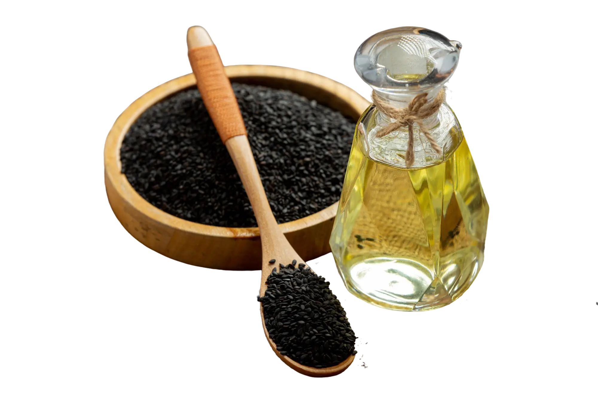
Sesame Oil: Is It Good for You?Sesame (Sesamum indicum L.) is one of the earliest human production and consumption oil crops in the family of Pedaliaceae, rape, soybean, and peanuts, known as China’s four major oil crops. Due to its highly aromatic odor and mellow flavor, sesame is extensively produced and well-liked. Sesame can be divided into three categories based on the color of the germplasm: white sesame, black sesame, and yellow sesame. Black and white sesame are the most prevalent and widely grown dominant species. Black sesame has strong growth ability, lodging resistance, and drought resistance, whereas white sesame has high oil content and good quality and has the largest planting area and distribution [1].Fat, protein, vitamins, minerals, and dietary fiber are all abundant in sesame seeds. Unsaturated fatty acids, fat-soluble vitamins, amino acids, and other nutrients are abundant in sesame oil, which is obtained through traditional oil production methods. Studies have found that sesame seeds contain 21.9% protein and 61.7% fat, and are rich in minerals. In addition to being rich in nutrients, sesame also contains many important functional components such as sesamin, sesamolin, sesamol, sesaminol, sesamolin phenol, and other lignan-like active ingredients. Each component of sesame has a different content depending on the extraction method and external growing conditions, e.g., hot-pressed sesame oil has a higher content of sesamol, sesamin, and total lignans than cold-pressed and refined sesame oil [1].Sesame oil is an aromatic oil extracted from sesame seeds and is a traditional product from the primary processing of sesame seeds, which can be used as edible oil. Linoleic and linolenic acids, as well as large quantities of biologically active compounds such as lignans, natural vitamin E, and phytosterols, are abundant in sesame oil. Sesame seed oil obtained by cold pressing has a high quality and nutritional content. The main unsaturated fatty acid in sesame oil is linoleic acid (46.9%), followed by oleic acid (37.4%). These fatty acids are essential fatty acids because they cannot be synthesized in the organism and must be obtained through the diet [1].Health Benefits of Sesame OilOsteoarthritis (OA) Sesame oil showed an attenuation of quadriceps muscle dysfunction in osteoarthritis (OA) rats. Lower muscle strength and muscle weakness are related to both increased interleukin (IL)-6 production and decreased citrate synthase (CS) activity in a number of animal illness models. Myosin heavy chain (MHC) typing change is one of the major causes of muscle weakness in various pathogenic situations. Lower muscular strength/muscle weakness in the quadriceps is related to decreased MHC IIa fiber in OA patients. In the present study, sesame oil effectively improved muscle dysfunction and elevated MHC IIa gene expression. It is likely that enhancing MHC IIa gene expression may be involved in sesame oil exerted attenuation of muscular dysfunction, at least partially [2].Sesame oil may improve quadriceps muscle dysfunction by inhibiting muscular oxidative stress during the initiation of OA. Increased oxidative stress in skeletal muscle is sufficient to cause muscular atrophy, according to genetic evidence. Elevated reactive oxygen species (ROS) generation can contribute to muscle dysfunction by oxidative damage, degradating contractile proteins, or activating calpain and ubiquitin proteolytic systems. Overproduction of ROS alters the fiber type and muscle function by regulating MHC gene expression. In addition, inhibiting endogenous antioxidant expression in mice results in significant loss of skeletal muscle mass and muscle weakness. In the present study, sesame oil may decrease joint pain by improving oxidative stress associated muscle dysfunction [2].Sesamin has been shown in studies to have anti-inflammatory properties. It is well recognized that TNF-α is crucial in the development of rheumatoid arthritis. Khansai et al., found that sesamin significantly reduced the mRNA expression of IL-6 and IL-1 in human primary synovial fibroblast cell lines, indicating that sesamin inhibited TNF-α-induced pro-inflammatory cytokine mRNA expression. The major sesamin metabolites found in human plasma after oral administration of sesamin are sesamin catechol conjugates. Catechol glucuronides exert anti-inflammatory effects through demyelination in macrophage-like J774.1 cells, thereby inhibiting the expression of interferon beta and inducible nitric oxide synthase. In murine macrophage-like J774.1 cells, it was found that SC1, one of CYP450's sesamin metabolites, has more potent anti-inflammatory properties than sesamin itself [1].Cardiovascular Disease and Lipid and Lipoprotein LevelsIt is generally known that lipids and lipoproteins play a causative role in cardiovascular disease (CVD). LDL-C and HDL-C are raised by dietary saturated fatty acids (SFA), which are present in milk, butter, cheese, cattle, lamb, hog, poultry, palm oil, and coconut oil. The increase in LDL-C is due to a decrease in hepatic LDL clearance and an increase in LDL production secondary to a decrease in hepatic LDL receptors. Sesame oil contains monounsaturated fatty acids (MUFA) which can decrease LDL-C by increasing hepatic LDL receptor activity [3].Not all meta-analyses, but the majority, have been unable to show that MUFA intake reduces cardiovascular events. However, one meta-analysis and the Nurses’ Health Study and Health Professionals Follow-Up Study, two very large observational studies, found that MUFA from plant sources was beneficial while MUFA from other sources was not protective from developing cardiovascular events [3].Metabolic syndromeInsulin resistance, dyslipidemia, hypertension, and abdominal obesity are all part of the metabolic syndrome, which raises the risk of type 2 diabetes and CVD. Inflammation and oxidative stress play a substantial role in the development of metabolic syndrome. Moreover, the metabolic syndrome is diagnosed by metabolic biomarkers such as increased triglycerides (TG) and decreased HDL, hypertension, obesity, insulin resistance, and elevated oxidative stress. These factors are the main causes of increase in mortality of patients with cardiovascular disease, type 2 diabetes, and strokes all over the world. Insulin sensitivity is increased by polyunsaturated fatty acids (PUFA), which have been shown to have various positive impacts on human health. Monounsaturated fatty acids (MUFA) reduce insulin resistance and TG through promoting fatty acid oxidation [4].Sesame oil is rich in MUFA, omega 6 Polyunsaturated fatty acids (PUFA) (83%-90%), such as oleic acid and linoleic acid, respectively. Sesame oil contains tocopherol, sesamin, sesamolin, polyphenols, phytosterols, flavonoids, and sesamol lignans that have anti-inflammatory and anti-mutagenic effects. Moreover, consuming this oil improves blood pressure, insulin levels, and fasting blood glucose (FBG). Vitamin B6, magnesium, calcium, copper, iron, and zinc are all found in sesame oil, which reduces blood pressure, hyperlipidemia, and lipid peroxidation by increasing enzymatic and nonenzymatic antioxidants. Sesamin, which is present in sesame oil, has anti-atherosclerotic effects that help to control blood pressure [4].The current systematic review and meta-analysis includes 12 clinical trials. They provided the evidence to show how consuming sesame oil improved metabolic biomarkers. Overall results showed sesame oil consumption significantly reduced FBG (-3.268 mg/ dl), and malondialdehyde (MDA; -4.847 mg/dl) compared to the control group. Also, HbA1C (-2.057%), systolic blood pressure (SBP; -2.679 mmHg), diastolic blood pressure (DBP; -1.981 mmHg), body weight (-0.346 kg), and body mass index (BMI; -0.385 kg/m2) were all significantly lower than baseline. However, no reduction effect was identified for insulin serum [4]. ReferencesWei P, Zhao F, Wang Z, Wang Q, Chai X, Hou G, Meng Q. Sesame (Sesamum indicum L.): A Comprehensive Review of Nutritional Value, Phytochemical Composition, Health Benefits, Development of Food, and Industrial Applications. Nutrients. 2022;14:1–26. https://www.mdpi.com/2072-6643/14/19/4079Hsu D, Chu P, Jou I. Enteral sesame oil therapeutically relieves disease severity in rat experimental osteoarthritis. Food Nutr Res. 2016;60:29807. https://www.ncbi.nlm.nih.gov/pmc/articles/PMC4816814/Feingold K. The Effect of Diet on Cardiovascular Disease and Lipid and Lipoprotein Levels. Endotext. 2021. https://www.ncbi.nlm.nih.gov/books/NBK570127/Atefi M, Entezari M, Vahedi H, Hassanzadeh A. The effects of sesame oil on metabolic biomarkers: a systematic review and meta-analysis of clinical trials. J Diabetes & Metab Disord. 2022;21:1065–80. https://www.ncbi.nlm.nih.gov/pmc/articles/PMC9167273/ 

THE POTENTIAL BENEFITS OF ZINC SUPPLEMENTS

THE POTENTIAL BENEFITS OF ZINC SUPPLEMENTS

March 18, 2025

THE POTENTIAL BENEFITS OF ZINC SUPPLEMENTSZinc is a trace element that is required for living organisms and their biological processes. Because the body cannot accumulate zinc, it is essential to take this element consistently in the diet [1].  In the human body, zinc is found in muscles (60%), bones (30%) and skin (5%). Zinc is involved in the activation of various enzymes and proteins, and it aids in the absorption of vitamins A, E, and folate. Low levels of zinc can be associated with an increased chance of developing infections and degenerative pathologies. Zinc also plays an important role in the psychosocial functioning of human behavior [2].Zinc deficiency defined by a plasmic zinc level below 60 µg/dl, exceeds the regulatory capacity of homeostatic mechanisms, clinical symptoms may arise. Zinc deficiency can occur due to inadequate intake, reduced absorption, increased losses, or increased demand. It can also occur due to genetic disorders such as Acrodermatitis enteropathica and sickle cell disease. Inadequate intakes as a result of a zinc-deficient diet or a phytate rich diet is the most common worldwide cause of zinc deficiency. Individuals most susceptible to zinc deficiency caused by inadequate intake are those with the greatest physiological demand. Due to age-related declines in absorption and poor diet, the elderly is particularly at risk [3]. Bullous-pustular dermatitis, alopecia, diarrhea, weight loss, intercurrent infections, and hypogonadism are all symptoms of severe zinc insufficiency in males. Unrecognized severe zinc deficiency is fatal. Growth retardation, delayed puberty, hypogonadism in males, rough skin, poor appetite, delayed wound healing, and abnormalities in gustation, olfaction, and night vision are all symptoms of moderate zinc deficiency. Oligospermia, weight loss, and hyperammonemia are all symptoms of mild zinc deficiency [3].THE BENEFITS OF ZINC SUPPLEMENTAcne VulgarisSince Michaelsson discovered zinc's beneficial effect on acne in a patient with acrodermatitis enteropathica, and subsequent studies demonstrated low serum zinc in acne patients, zinc has been widely used both topically and systemically for the treatment of acne vulgaris. Oral zinc sulfate is reportedly more effective in the treatment of severe acne than for the treatment of mild to moderate acne but nausea, vomiting, and diarrhea occur frequently. Oral zinc gluconate has also been reported to be effective in the treatment of inflammatory acne, but the initial loading dose is ineffective. However, acne treatment with zinc salts appears to be equal or less effective compared with systemic tetracyclines (minocycline, oxytetracycline). The exact mechanism of zinc in acne treatment remains poorly elucidated and is considered to act directly on microbial inflammatory equilibrium and facilitate antibiotic absorption when used in combination. Another mechanism for zinc's acne benefit is its antiandrogenic action, which suppresses sebum production [4].AlopeciaAndrogenetic alopecia is a prevalent condition in males over the age of 20, with an estimated 90% of males over the age of 20 experiencing some degree of frontal recession. The mainstay of treatment is minoxidil and finasteride, as well as surgical methods like hair transplantation. Zinc has been discovered to have antiandrogen properties and to alter the activities of 5𝛼-reductase types 1 and 2 [4]. ImmunityZinc is a trace element that is required for the activity of several enzymes and transcription factors in humans. It plays a key role in regulating the function of both the adaptive and the innate immune system. Dietary sources of zinc are animal products such as meat, fish, eggs, and dairy, but it is also contained in whole grains, nuts, and legumes. When compared to zinc obtained from plant products, zinc from animal sources has a higher bioavailability. Phytate, some dietary fibers, and lignin are plant ligands that chelate zinc and prevent it from being absorbed [5].Zinc’s effect on the immune system is complex; it can promote and inhibit various immunological activities to achieve a proper balance of pro and anti-inflammatory effects via a variety of methods. A correct intake of zinc is essential to limit the overproduction of inflammatory cytokines: in vitro and human studies show that zinc deficiency is associated with an increased inflammatory response and excessive release of pro inflammatory cytokines such as IL-2, IL-6, and TNF-alfa, regulated through the NF-κB signaling pathway. Zinc also enhances the number of inducible regulatory T cells. Another important role played by zinc is the maintenance of membrane barrier integrity, which is essential in the pulmonary and intestinal epithelia that constitute the first barrier to protect the organism from pathogens. Zinc supplementation has also been proven to have a direct antiviral effect on RSV, Dengue virus, and coronaviruses, as well as reducing oxidative stress and minimizing the duration of cold symptoms in adults. Lastly, some authors suggested that combining chloroquine with zinc would increase the toxicity of chloroquine against viruses [5].Zinc supplementation has also been investigated against acute lower respiratory infection. Two studies found that zinc therapy decreased infection episodes and increased recovery rates from illness and fever, however the latter study's effect was only significant in boys. Lower respiratory infection may be caused by a variety of bacteria or viruses, the nature of which was not explored in these studies. Hence it can be concluded that zinc treatment reduces the symptoms, but there is no evidence that zinc has an effect on the immune response to the underlying infections based on these findings. A pooled analysis of four trials in which continuous supplementation was investigated confirmed that zinc is efficient for the prevention of pneumonia. In this study, zinc supplementation reduced the incidence of pneumonia in children in developing countries by 41%. Pneumonia is a major cause of childhood mortality; it accounts for approximately 20% of childhood deaths in developing countries, making zinc supplementation a promising approach for a significant reduction in childhood mortality. Furthermore, a recent study suggests that zinc may be beneficial to the elderly. Zinc supplementation was found to be associated with a lower risk of pneumonia in nursing home residents, suggesting that zinc supplementation could be used to prevent pneumonia in the elderly [6].Wound HealingZinc, a trace mineral, is found to be in low concentration in tissues and across cell membranes in post-neurosurgical wound-healing patients. As such, zinc is firmly regulated through gene transcription rule, ion carriers, cellular homeostasis, and extracellular supplies. Extracellular vesicles contain a modest amount of zinc during physiological processes. Zinc is normally taken up by the zinc transporter protein (ZIP) in intracellular vesicles. The cytosol contains free zinc ions, which have been identified as secondary messengers capable of targeting proteins to regulate a variety of chemical and physiological pathways. Therefore, the availability of zinc and its regulation are essential components of cellular physiology [7].Zinc shortage has been blamed for delays in wound healing. Zinc deficiency plays a role in inflammation by increasing the inflammatory response and causing damage to the host tissue. Post-neurosurgical and severely ill patients, patients with severe burn injury, hypodermic sore, insignificant surgery, and pressure ulcers have all received zinc supplements [7].  INTERACTIONZinc–IronStudies showed that high concentrations of iron can have a negative effect on zinc absorption in human adults when zinc and iron are given in solution and on an empty stomach. It has been suggested that suppression of zinc absorption by iron occurs when given in an aqueous medium because of a competition for common nonspecific pathways. This suppression cannot occur when iron and zinc are given during a meal because zinc can be absorbed via an alternate pathway with the aid of ligands formed during protein digestion [8].Zinc–VitaminsFinally, the interaction of zinc with some vitamins (A, D, and E) is intriguing because zinc influences the vitamins' trans-porters, such as retinol-binding protein for vitamin A and tocopherol for vitamin E. The interaction between zinc and vitamin D is suggestive because zinc participates in the constitution of vitamin D (especially D3 isoform) receptor DNA binding domain through two zinc finger-like motifs, favoring the re-absorption of calcium at kidney level and phosphorus at intestinal level. As a result, the interaction of zinc and vitamin D is critical for the proper functioning of a variety of organs and tissues, including the brain and bone [8].Zinc–CalciumAlthough different mechanisms of zinc and calcium absorption in small intestine have been proposed through the binding with prostaglandin E and vitamin D, respectively, experiments in rats have however shown that calcium may interfere in intestinal zinc absorption because a competition between zinc and calcium for the same transcellular transporting carriers on the mem-brane surface occurs [8]. REFERENCESSanna A, Firinu D, Zavattari P, Valera P. Zinc Status and Autoimmunity: A Systematic Review and Meta-Analysis. Nutrients. 2018;10:1–17. https://www.ncbi.nlm.nih.gov/pmc/articles/PMC5793296/Uwitonze AM, Ojeh N, Murererehe J, Atfi A, Razzaque MS. Zinc Adequacy Is Essential for the Maintenance of Optimal Oral Health. Nutrients. 2020;12:1–14. https://www.ncbi.nlm.nih.gov/pmc/articles/PMC7230687/Kogan S, Sood A, Garnick MS. Zinc and Wound Healing: A Review of Zinc Physiology and Clinical Applications. Wounds. 2017;29:102–6. https://www.hmpgloballearningnetwork.com/site/wounds/article/zinc-and-wound-healing-review-zinc-physiology-and-clinical-applicationsGupta M, Mahajan VK, Mehta KS, Chauhan PS. Zinc therapy in dermatology: a review. Dermatol Res Pract. 2014;2014:. https://www.ncbi.nlm.nih.gov/pmc/articles/PMC4120804/Pecora F, Persico F, Argentiero A, Neglia C, Esposito S. The Role of Micronutrients in Support of the Immune Response against Viral Infections. Nutrients. 2020;12:1–45. https://www.ncbi.nlm.nih.gov/pmc/Overbeck S, Rink L, Haase H. Modulating the immune response by oral zinc supplementation: a single approach for multiple diseases. Arch Immunol Ther Exp. 2008;56:15–30. https://ww.ncbi.nlm.nih.gov/pmc/articles/PMC7079749/Adjepong D, Jahangir S, Malik B. The Effect of Zinc on Post-Neurosurgical Wound Healing: A Review. Cureus. 2020;12:1–7. https://www.ncbi.nlm.nih.gov/pmc/articles/PMC7039353/Mocchegiani E, Romeo J, Malavolta M, Costarelli L, Giacconi R, Diaz LE, Marcos A. Zinc: dietary intake and impact of supplementation on immune function in elderly. Age (Dordr). 2013;35:839–60. https://www.ncbi.nlm.nih.gov/pmc/articles/PMC3636409/

The benefits of “Ginkgo Biloba Extract”

The benefits of “Ginkgo Biloba Extract”

March 16, 2025

The benefits of “Ginkgo Biloba Extract”Ginkgo biloba is one of the oldest living species on the planet. The leaves and seeds of this plant have been used for medicinal purposes in China for centuries, initially being used for asthma and problems in the digestive system. In Europe and the USA, they have been sold since the sixties, and now they represent one of the most popular phytotherapeutic products in the world [1].In the early 1970s, Dr. Willmar Schwabe Pharmaceuticals (Karlsruhe, Germany) effectively developed a method for the extraction and standardization of ginkgo biloba extract preparation and produced highly concentrated and stable extract from ginkgo biloba leaves. The standardized extract of ginkgo biloba leaves contains 6% terpenoids (in which 3.1% are ginkgolides A, B, C, and J and 2.9% is bilobalide), 24% flavonoid glycosides (containing quercetin, kaempferol, isorhamnetin etc.), and 5–10% organic acids [2].The active compounds for ginkgo biloba extract is flavonoids and terpenoids. The content of flavonoids promotes blood circulation, especially cerebral blood circulation. Therefore, it is used in disorders such as Alzheimer’s disease and memory loss that are caused by decreased blood flow. In addition, the extract also has antioxidant [3] and neuroprotective properties [2]. Ginkgo Biloba Extracts PropertiesDementia and Alzheimer’s diseaseThe rapid aging of the global population is resulting in an increasing prevalence of mild cognitive impairment, dementia, and Alzheimer's disease. Age is a key risk factor for these diseases; after the age of 65 years, the prevalence of dementia doubles every five years. It is higher among women than men, largely because women tend to live longer [4].There are studies performed that the positive effect of ginkgo biloba extract has been reported on Alzheimer’s disease, memory enhancement, dementia of vascular origin, and cognitive disorders. The extract modifies the cerebral blood flow and may help to reduce fatigue and inattention [2]. But some studies of ginkgo biloba extract have not been demonstrated to prevent the progression of dementia. However, several studies showed that ginkgo biloba extract encourage efficacy in terms of improvement in cognition, behavior, and ability to maintain activities of daily living in patients with dementia or Alzheimer's disease, as well as reducing caregiver burden [4]. Age-related macular degeneration (AMD)Age-related macular degeneration (AMD) is a condition affecting the central area of the retina (the back of the eye). The retina can deteriorate with age and some people get lesions that lead to loss of central vision. Vascular factors and oxidative damage are two potential mechanisms in the pathology of the disease. Ginkgo biloba extract contains two constituents (flavonoids and terpenoids) which have antioxidant properties. It is believed these may help to slow down the progression of AMD through several mechanisms of action including increased blood flow, inhibiting platelet-activating factor, and prevention of membrane damage caused by free radicals[5]. GlaucomaGlaucoma is the leading cause of irreversible visual impairment and blindness. Although the mechanism of glaucoma is still largely unknown, oxidative stress, optic nerve ischemia, and neuroinflammation were found to be a certain role in the development of glaucomatous optic nerve degeneration. It has been suggested that ginkgo biloba extract may protect tissue against free radical damage like other antioxidants such as vitamin C and E. However, unlike the others, the extract acts at the level of organelles by stabilizing the mitochondria due to abnormal mitochondrial changes can lead to glaucoma [6].   Ginkgo biloba extract was also found to have vasodilatory properties that could improve coronary and peripheral circulation that could improve blood viscosity [6]. Moreover, it was associated with positive effects on the improvement of deteriorated visual field in patients with open-angle glaucoma [7]. Due to its antioxidant, vasoregulatory, and anti-inflammatory benefits, ginkgo biloba extract is considered a neuroprotective agent and improve the vision of people with glaucoma [6].Oxidative stressOxidative stress is caused by destructive and progressive modifications in one or more body tissues, leading to dysfunction of organs, premature aging, and sometimes disease and death. Stress exposure, radiation, infections, and smoke exposure cause significant damage, inducing alterations in DNA, can lead to several oxidative disorders such as cardiovascular diseases and cancer. It is possible that the usage of ginkgo biloba extract may delay this process [1].Ginkgo biloba extract has antioxidant properties since it regulates the expression of antioxidant enzymes positively and reduces reactive oxygen and nitrogen species. It also exhibits anti-inflammatory properties, inhibits the expression of pro-inflammatory cytokines, such as IL-1, IL-6, and TNF-α so it minimizes the risk factors for diseases that are caused by the oxidative stress process [1].Warnings, Interactions, and Adverse Effects of Ginkgo Biloba ExtractThe most important clinical problem with ginkgo biloba is caused by its inhibition of the platelet-activating factor, so the use of ginkgo in conjunction with warfarin, aspirin, or other antiplatelet agents should be consulted with a specialist. Ginkgo biloba is generally well tolerated, with side effects being rare, usually mild, and including nausea, vomiting, diarrhea, headaches, dizziness, palpitations, restlessness, weakness, or skin rashes. Although no studies have been performed to support any restrictions on the use of ginkgo during pregnancy or lactation, it seems careful not to administer ginkgo in the absence of any data[8]. ReferencesAchete de Souza G, de Marqui SV, Matias JN, Guiguer EL, Barbalho SM. Effects of Ginkgo biloba on Diseases Related to Oxidative Stress. Planta Medica. 2020;86:376–86. เข้าถึงจาก: https://www.thieme-connect.com/products/ejournals/abstract/10.1055/a-1109-3405Singh SK, Srivastav S, Castellani RJ, Plascencia-Villa G, Perry G. Neuroprotective and Antioxidant Effect of Ginkgo Biloba Extract Against AD and Other Neurological Disorders. Neurotherapeutics. 2019;16:666–74. เข้าถึงจาก: https://www.ncbi.nlm.nih.gov/pmc/articles/PMC6694352/Sellami M, Slimeni O, Pokrywka A, Kuvačić G, D Hayes L, Milic M, Padulo J. Herbal medicine for sports: a review. J Int Soc Sports Nutr. 2018;15:1–14. เข้าถึงจาก: https://www.ncbi.nlm.nih.gov/pmc/articles/PMC5856322/Kandiah N, Ong PA, Yuda T, Ng LL, Mamun K, Merchant RA, et al. Treatment of dementia and mild cognitive impairment with or without cerebrovascular disease: Expert consensus on the use of Ginkgo biloba extract, EGb 761®. CNS Neurosci Ther. 2018;25:288–98. เข้าถึงจาก: https://www.ncbi.nlm.nih.gov/pmc/articles/PMC6488894/Evans J. Ginkgo biloba extract for age-related macular degeneration (Review). Cochrane Database Syst Rev. 2013:1–15. เข้าถึงจาก: https://www.ncbi.nlm.nih.gov/pmc/articles/PMC7061350/Ige M, Liu J. Herbal Medicines in Glaucoma Treatment. Yale J Biol Med. 2020;93:347–53. เข้าถึงจาก: https://www.ncbi.nlm.nih.gov/pmc/articles/PMC7309662/Bungau S, Abdel-Daim MM, Tit DM, Ghanem E, Sato S, Maruyama-Inoue M, et al. Health Benefits of Polyphenols and Carotenoids in Age-Related Eye Diseases (Review). Oxid Med Cell Longev. 2019:1–22. เข้าถึงจาก: https://www.ncbi.nlm.nih.gov/pmc/articles/PMC6390265/Sierpina VS, Wollschlaeger B, Blumenthal M. Ginkgo Biloba. Am Fam Physician. 2003;68:923–6. เข้าถึงจาก: https://www.aafp.org/afp/2003/0901/p923.html

Pomegranate extract

Pomegranate extract

March 14, 2025

POMEGRANATE EXTRACTThe pomegranate (Punica granatum L.), belonging to Punica L. genus, Punicaceae family, is an ancient fruit native to Central Asia in regions spanning from Iran and Turkmenistan to northern India as well as in the Mediterranean area and the Middle East. Pomegranate and its components have been shown in studies conducted over the last several decades to have potent anti-oxidative and anti-inflammatory capabilities. In addition to in vivo and in vitro studies showed that pomegranate exhibits anti-hypertensive and anti-proliferative properties [1].Pomegranate fruit is abundant in flavonoids, proanthocyanidins, and natural polyphenols. It is frequently used as nutritional food, for medicinal purposes, health promotion, and strong anti-oxidant action. As pomegranate seeds include vitamin C, vitamin K, folic acid, and bioactive chemicals rich in polyphenols, they have been shown to scavenge reactive oxygen species (ROS). Furthermore, the pomegranate fruit peel was estimated to primarily contain 25 - 28% of the polyphenols with gallic acid and tannins for scavenging ROS, while the pomegranate fruit pericarp mainly contains potent bioactive compounds, such as gallagic, ellagic acid, ellagitannins, punicalagin, anthocyanins delphinidin, pelargonidin, and luteolin [2].  The Benefits of Pomegranate ExtractVasculoprotective Effects  The presence of hydrolyzable tannins (ellagitannins and gallotannins), pomegranate derivative ellagic acid, or their common metabolites urolithins was hypothesized to be the reason for the protective benefits [1].In hypertensive patients, daily consumption of pomegranate juice for 2 weeks reduced the activity of angiotensin-converting enzyme (ACE) by 36% as well as diminished systolic blood pressure by 5%. The group also reported that patients with carotid artery stenosis significantly decreased their blood pressure, low-density lipoprotein (LDL) oxidation, and common carotid intima-media thickness by consuming pomegranate juice over 3 years. In a cohort of 51 healthy women, pomegranate juice drinking for four weeks significantly lowered blood pressure (without significantly changing serum ACE activity). A subsequent study involving 21 hypertensive patients revealed that pomegranate juice consumption effectively lowered both systolic and diastolic blood pressure [1]. Inflammatory DiseaseConsuming pomegranates has a generally positive effect on patients with chronic inflammatory disorders. In patients with hypertension or metabolic syndrome, as well as those undergoing dialysis, pomegranate juice appears to offer promising hypotensive properties. It also resulted in a slight amelioration of lipid profiles in patients with cardiovascular disease (CVD), as pomegranate intake elevated endogenous levels of high-density lipoprotein (HDL) cholesterol and reduced triglyceride (TG) levels. However, several studies have been unable to confirm pomegranate’s TG and cholesterol lowering effect. With regards to risk factors for CVD, one dose of 150 mL of pomegranate juice per day did not influence the level of circulating soluble adhesion molecules or indicators of atherosclerosis and subclinical coronary heart disease (CHD) in hypertensive people. Long-term consumption (100 mL per day for a year) or a higher intake of juice (500 mL per day) were both used to demonstrate the beneficial effects of pomegranate on CVD [3].Several groups have studied the effects of pomegranate on the prevention and amelioration of atherosclerosis and other CVD symptoms. In a rat model of the metabolic syndrome, de Nigris et al. (2007) found that supplementation with pomegranate juice or pomegranate fruit extract decreased the expression of vascular inflammatory markers and transforming growth factor β-1 (TGFβ-1), and, likewise, elevated endothelial nitric oxide synthase (eNOS) levels. Additionally, Labsi et al. (2016) demonstrated that Swiss albino mice treated intraperitoneally with pomegranate peel extract for two months after the induction of echinococcosis significantly reduced the nitric oxide (NO) and TNF-α levels [3].Anti-oxidantReactive oxygen species (ROS) are a by-product of the natural metabolism of oxygen in mammalian bodies, and are neutralized by the anti-oxidant system. Increased ROS production due to exposure to toxic chemicals and xenobiotics leads to an imbalance between the production of ROS and their elimination by anti-oxidant systems. The increased ROS attack cell membranes and damage biomolecules such as proteins and DNA [4].Natural anti-oxidants found in pomegranates, such as anthocyanins, catechins, quercetin, gallotannins, ellagitannins, ellagic, ferulic, and gallic acid, are abundant and show promise as anti-oxidants. The most prevalent polyphenols in the peel are ellagitannins (punicalagin and its derivatives), which are known for their strong anti-oxidant properties [4].Pomegranate is a rich source of polyphenols and several studies have shown the beneficial role of both pomegranate and its bioactives on reducing oxidative stress and lipid peroxidation through the direct neutralization of ROS, upregulating anti-oxidant enzymes, and modulating transcription factors such as nuclear factor κB (NF-κB) or peroxisome proliferator-activated receptor g (PPARg), among others. The primary bioactive components of pomegranate fruit extract that support skin health are ellagic acid and punicalagin, which have anti-oxidant and anti-inflammatory properties as well as the ability to inhibit the tyrosinase enzyme. Additionally, pomegranate extract stimulates type I procollagen synthesis and inhibits MMP-1 (collagenase) production by dermal fibroblasts [5].As confirmed by the reduced histopathological damage, the pomegranate-peel extract meaningfully restored the liver markers. This is consistent with the results in, where the authors revealed that pomegranate’s anti-oxidant properties could reduce oxidative-stress-induced liver injury. It has been discovered that pomegranate phenolic compounds contain anti-oxidant and free-radical-scavenging characteristics, and they also considerably enhanced the kidney weight to body weight ratio [4].  ReferencesWang D, Özen C, Abu-Reidah I, Chigurupati S, Patra J, Horbanczuk J, et al. Vasculoprotective Effects of Pomegranate (Punica granatum L.). Frontiers in Pharmacology. 2018 [cited 2022 November 24]; 9: 1-15. Available form: https://www.frontiersin.org/articles/10.3389/fphar.2018.00544/full#B108Singh M, Lee K, Vinayagam R, Kang S. Antioxidant and Antibacterial Profiling of Pomegranate-pericarp Extract Functionalized-zinc Oxide Nanocomposite. Biotechnology and Bioprocess Engineering. 2021 [cited 2022 November 29]; 26: 728-37. Available form: https://www.ncbi.nlm.nih.gov/pmc/articles/PMC8548265/Danesi F, Ferguson L. Could Pomegranate Juice Help in the Control of Inflammatory Diseases? Nutrients. 2017 [cited 2022 December 6]; 9: 1-23. Available form: https://www.mdpi.com/2072-6643/9/9/958Sayed S, Alotaibi S, El-Shehawi A, Hassan M, Shukry M, Alkafafy M, et al. The Anti-Inflammatory, Anti-Apoptotic, and Antioxidant Effects of a Pomegranate-Peel Extract against Acrylamide-Induced Hepatotoxicity in Rats. Life (Basel). 2022 [cited 2022 December 6]; 12: 1-16. Available form: https://www.mdpi.com/2075-1729/12/2/224Quiles J, Cabrera M, Jones J, Tsapekos M, Caturla N. In Vitro Determination of the Skin Anti-Aging Potential of Four-Component Plant-Based Ingredient. Molecules. 2022 [cited 2022 December 6]; 27: 1-23. Available form: https://www.mdpi.com/1420-3049/27/22/8101

WHAT ARE THE BENEFITS OF GRAPE SEED EXTRACT?

WHAT ARE THE BENEFITS OF GRAPE SEED EXTRACT?

March 13, 2025

WHAT ARE THE BENEFITS OF GRAPE SEED EXTRACT?Grape seed extract (GSE) is having abundant source of polyphenols. Polyphenols and flavonoids present in the GSE have been shown remarkable interest based on positive reports of their antioxidant properties and ability to serve as free radical scavengers. In comparison to other well-known antioxidants (such vitamin C, vitamin E, and β-carotene), grape seed polyphenols have a higher antioxidant activity. Along with its antioxidant property, it also has several enzymes that catalyze the release of histamine in allergic reactions and inflammatory conditions. The amount of oil present in grape seed depends on the variety of the grape (usual range 10 - 16% of dry weight). In addition, grape seed oil has a high amount of unsaturated fatty acids, including α-linolenic acid (ω - 3) and g-linolenic acid (ω - 6) from 85 to 90%. These fatty acids are related to a reduction of cardiovascular disease, cancer, hypertension, and autoimmune disorders. An oil containing high amount of linoleic acid results in the reduction of total blood cholesterol and low-density lipoprotein (LDL) cholesterol. Due to the fact that LDL cholesterol is responsible for the formation of arteriosclerosis, the application of grapeseed oil has a good impact on the decrease of arteriosclerosis. Linoleic, linolenic, oleic, and palmitic acids are among the fatty acids present in GSE and play a significant role in lipid metabolism. The major fatty acid in GSE is linoleic acid followed by oleic acid and palmitic acid. The seeds and peels of grapes also contains considerable portion of dietary fiber that lowers the risks of colon cancer, heart disease, diabetes and obesity [1]. The Benefits of Grape Seed ExtractWound Healing Activity The most effective growth factor in the process of wound healing is vascular endothelial growth factor (VEGF). GSE contains polyphenyl phenolic bioflavonoids, proanthocyanidins which accelerates the process of wound healing. Proanthocyanidins specifically induces the VEGF expression in human keratinocytes cells responsible for wound healing. A phenolic component found in GSE has been shown in some epidemiological studies to decreases the endothelial contraction of vessels, activate nitric oxide synthesis, regulate platelet aggregation and prevent LDL cholesterol oxidation [1].   Anti-inflammationPro-inflammatory cytokines are activated through downstream cascades during persistent inflammation in wounds. These cytokines include tumor necrosis factor (TNF), interleukin (IL)-1, IL-6, and IL-17. There are studies that have demonstrated the anti-inflammatory potential of GSE on murine macrophage RAW264.7 cells. According to these studies, GSE has a significant potential to reduce the production of inflammatory markers including tumor necrosis factor-α (TNFα), inducible isoform of nitric oxide synthase (iNOS), IL-6, and nitric oxide (NO). This is accomplished by inhibiting the phosphorylation p38, ERK1/2, JNK, and NF-κB signaling pathways through a synergistic interaction of the phenolic compounds and flavonoids present in the GSE [2].Antioxidant PotentialIt is well known that plant polyphenols, particularly GSE, have antioxidant property. These bioactive molecules exert their beneficial effects by neutralizing reactive oxygen species (ROS) and chelating pro-oxidative metal ions [1, 3]. Polyphenols may play a key role in the prevention of degenerative processes, such as cancer, diabetes, chronic inflammation and aging. Additionally, polyphenolic compounds inhibit the oxidation of LDL and prevent platelet aggregation, decreasing the development of cardiovascular and coronary diseases [3].ImmunityNumerous studies have demonstrated the immunity function of GSE. The immunity may be influenced by oxidative stress, and the improved antioxidant function may enhance their immune function in poultry. It has been showed in pigs that polyphenols may boost immune function by suppressing inflammation via nuclear factor-kappaB and nuclear factor-2-dominant pathways in the small intestine. Interferon-γ (IFN-γ) serves as an important regulator in the activation of lymphocytes and monocytes and serum IL-2 promotes the proliferation of activated natural killer cells, B lymphocytes, T lymphocytes, and antibody production. Complement4 (C4) is a crucial component of the body's immunological defense system and plays a significant role in immune response. According to the current findings, the supplementation of GSE improved serum C4, IL-2, and INF-γ, indicating GSE could enhance immune response by regulating antibodies, complements, and cytokines [4].Cardiovascular DiseasesIn the presence of risk factors for cardiovascular diseases (CVDs), including hypertension, diabetes mellitus, dyslipidemia, and smoking, increased generation of ROS contributes to endothelial dysfunction. For example, elevated ROS levels in hypertensive patients leads to reduced vascular bioavailability of NO. Hence, antioxidants that improve oxidative stress status are expected to promote vascular health, thereby preventing CVDs. Several studies involving rodents and humans with hypertension or CVDs have demonstrated how antioxidants, such as the vitamins C and E, genistein, and polyphenols, improve vascular endothelial function and lower blood pressure by reduction in ROS levels. A type of polyphenols called proanthocyanidin has potent antioxidant properties. Proanthocyanidin has greater antioxidant activity than vitamins C and E, β-carotene, or monomeric flavanol, including (+)-catechin. Furthermore, grape seed extracts containing 39-73% proanthocyanidin have also been shown to have strong antioxidant potency [5].The in vitro and in vivo studies show a relation between presence of antioxidant phenolics and the reduction of oxidized LDL particles, and intake of diets high in phenolic antioxidants decreased the incidence of heart disease. The ability of the polyphenols in GSE avert radical oxidation of the polyunsaturated fatty acids of LDL, which occurs frequently through oxidative modification of the apoprotein toward an atherogenic form, has a direct follow up on the prevention of CVDs [1].Skin AgingGrape seed has the ability to improve skin health and reduce skin aging by: (a) functioning as antioxidants and encouraging the Nrf2 pathway to generate other antioxidants, (b) promoting collagen, elastin, and TIMP 1 while, at the same time, inhibiting MMPs and the enzyme elastase, (c) inhibiting inflammatory molecules such as the interleukins and cyclo-oxygenase (COX) compounds, and the inhibition or counteracting of ROS formation, (d) inhibiting NF kappaB and AP-1, (e) stimulating SIRT 1 gene expression (the anti-aging biomarker), (f) binding of the potent androgen, 5α-DHT, and inhibiting the 5α-reductase enzyme in fibroblasts, and (g) binding to estrogen receptor β in the keratinocytes in the epidermis and fibroblasts in the dermis [6]. ReferencesGupta M, Dey S, Marbaniang D, Pal P, Ray S, Mazumder B. Grape seed extract: having a potential health benefits. Journal of Food Science and Technology. 2020 [cited 2022 October 27]; 57: 1205-15. Available form: https://www.ncbi.nlm.nih.gov/pmc/articles/PMC7054588/ Ajit A, Vishnu A, Varkey P. Incorporation of grape seed extract towards wound care product development. 3 Biotech. 2021 [cited 2022 October 27]; 11: 1-10. Available form: https://www.ncbi.nlm.nih.gov/pmc/articles/PMC8113434/Szabó É, Marosvölgyi T, Szilágyi G, Kőrösi L, Schmidt J, Csepregi K, et al. Correlations between Total Antioxidant Capacity, Polyphenol and Fatty Acid Content of Native Grape Seed and Pomace of Four Different Grape Varieties in Hungary. Antioxidants (Basel). 2021 [cited 2022 October 27]; 10: 1-12. Available form: https://www.mdpi.com/2076-3921/10/7/1101Ao X, Kim I. Effects of grape seed extract on performance, immunity, antioxidant capacity, and meat quality in Pekin ducks. Poultry Science. 2020 [cited 2022 October 27]; 99: 2078-86. Available form: https://www.sciencedirect.com/science/article/pii/S0032579119580580?via%3DihubOdai T, Terauchi M, Kato K, Hirose A, Miyasaka N. Effects of Grape Seed Proanthocyanidin Extract on Vascular Endothelial Function in Participants with Prehypertension: A Randomized, Double-Blind, Placebo-Controlled Study. Nutrients. 2019 [cited 2022 October 27]; 11: 1-12. Available form: https://www.mdpi.com/2072-6643/11/12/2844#B20-nutrients-11-02844 David L, Virginia A, Edwin D. Enhancing Skin Health: By Oral Administration of Natural Compounds and Minerals with Implications to the Dermal Microbiome. International Journal of Molecular Sciences. 2018 [cited 2022 January 31]; 19: 1-35. Available form: https://www.mdpi.com/1422-0067/19/10/3059